Figure 4.

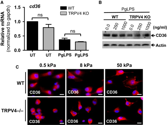
Reduced foam cell formation in TRPV4 KO cells is independent of expression of CD36. (A) qRT‐PCR analysis was performed to determine levels of CD36 in WT and TRPV4 KO MRMs using a TaqMan gene Expression Assay. All Ct values were normalized to gapdh levels. The experiment was repeated three times in quadruplicate. ns, not significant; Student's t‐test. (B) Representative immunoblots from three independent experiments show expression of CD36 and actin proteins in whole cell extract from WT and TRPV4 KO MRMs with or without PgLPS treatment. (C) WT and TRPV4−/− MRMs were maintained on various stiffness collagen‐coated (10 μg/mL) polyacrylamide gels (0.5, 8, and 50 kPa) for 48 h, and then stained with anti‐CD36 IgG (red color). Representative immunofluorescence images from three independent experiments are shown (original magnification, 40×).
