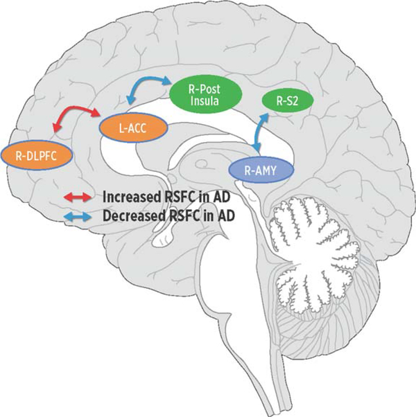
Fig. 2.
Regions in networks demonstrating increased (red arrows) or decreased (blue arrows) resting-state functional connectivity (RSFC) in Alzheimer’s disease (AD). Relative to controls, people with AD demonstrated increased RSFC between the cognitive (dlPFC) and affective (ACC) regions in the medial pain system while conversely people with AD displayed decreased RSFC (ACC) regions in the medial network and sensory (pINS) network and between the sensory (S2) network and the descending modulatory (AMY) network. dlPFC, dorsolateral prefrontal cortex; ACC, anterior cingulate cortex; pINS, posterior insula; S2, secondary somatosensory cortex; AMY, amygdala.

