
Fig. 3.
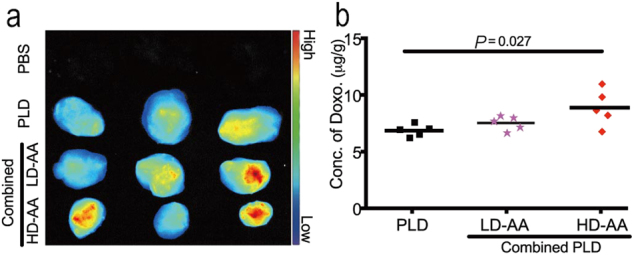
Doxorubicin accumulation in SW620 xenograft tumor. (a) Ex vivo fluorescence imaging. The intensity of doxorubicin in the HD-AA combined group was significantly increased compared with mono-PLD treatment. (b) Concentration of doxorubicin quantified by HPLC-MS/MS. The intratumor level of doxorubicin was significantly increased by combination with high-dosage AA (4 mg/kg, red diamond) compared with mono-PLD treatment (black square, P = 0.005), whereas low-dosage AA (1 mg/kg, pink star) failed to enhance intratumor exposure of doxorubicin