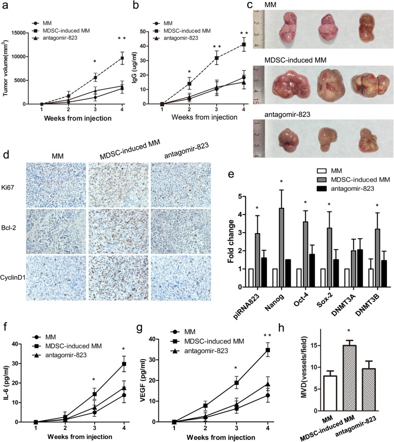
Fig. 5.
MDSCs enhance tumor growth and stemness through the piRNA-823 pathway in RPMI8226-xenografted mice. a-c MM cells, MDSCs-induced MM cells, and MDSCs-induced AS-MM cells were subcutaneously injected into nude mice (n = 3 for each group). Tumor volumes and IgG levels was monitored weekly. Data are presented as mean tumor volumes ± SD. * P < 0.05, ** P < 0.01. d Representative images of immunohistochemical staining for Ki67, Bcl-2, and CyclinD1 in tumor xenografts are shown (original magnification × 400). e Relative expression levels of the selected makers in tumor xenografts were detected using qRT-PCR. Values represent the means of three different experiments compared to the control ± SD. * P < 0.05. f and g Circulating IL-6 and VEGF levels in nude mice were detected using ELISAs. Significantly higher circulating IL-6 and VEGF levels were detected in the MDSC-induced MM group than in the other 2 groups. * P < 0.05, ** P < 0.01. h The quantitative analysis of microvessel density (MVD) was performed by counting the number of stained vessels in each group per high-power field (HPF) under the microscope. Data are presented as the means ± SEM of five HPFs. * P < 0.05

