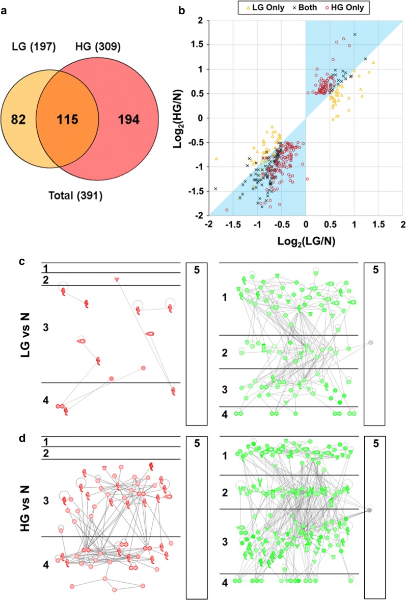
Fig. 3.
Comparison of protein groups differentially expressed in LG and HG PCa, compared with N samples. a Venn diagram of protein groups differentially expressed in LG and HG PCa, compared with N samples. A total of 82 DEPs were LG only, 194 were HG only, and 115 were shared by both LG and HG. b Scatter plot for the comparison of log2(LG/N) and log2(HG/N) ratios for the 82 LG-only, 115 shared, and 194 HG-only DEPs. The cyan shade covers the area where the absolute values of log2(LG/N) are less than those of log2(HG/N), i.e., the changes in the LG group are less remarkable than those in the HG group. c Putative networks of direct PPIs for proteins significantly upregulated (left) or downregulated (right) in LG PCa, compared with N samples. The five subcellular localization layers are (1) extracellular space, (2) plasma membrane, (3) cytoplasm, (4) nucleus, and (5) others. More detailed information is shown in Supplemental Figure S3. d Putative networks of direct PPIs for proteins significantly upregulated (left) or downregulated (right) in HG PCa, compared with N samples. The subcellular localization layers are the same for the panel C. More detailed information is shown in Supplemental Figure S4. In the figure, the abbreviations N, LG, and HG stand for normal prostate, low-grade PCa, and high-grade PCa, respectively

