Fig. 1.
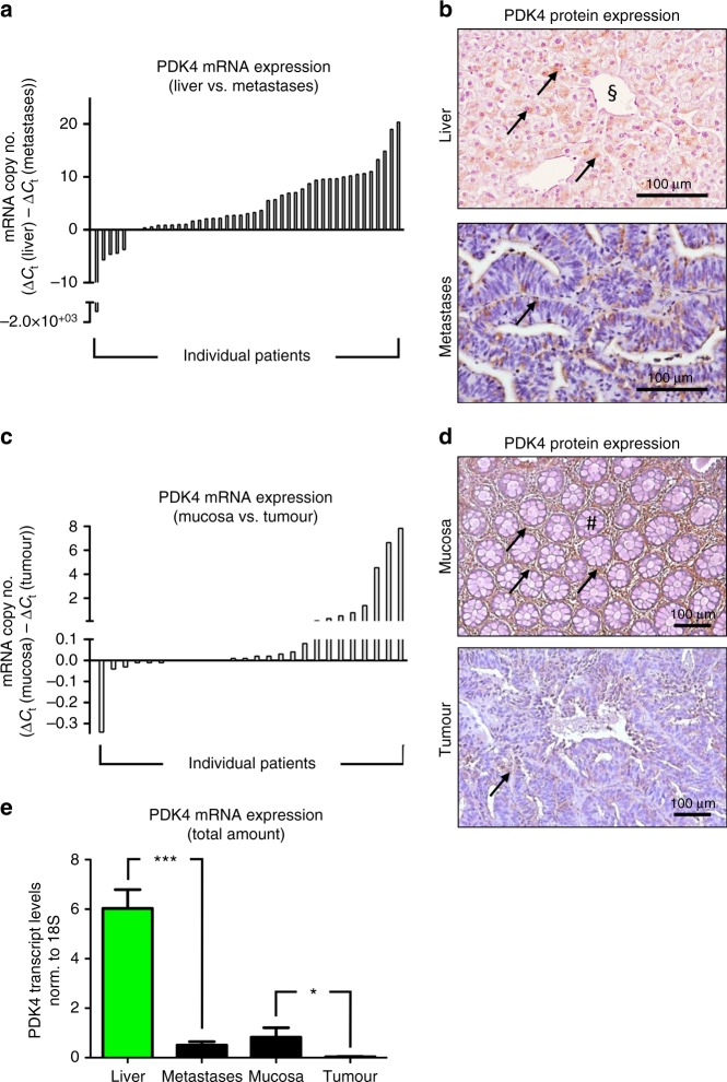
PDK4 expression in CRLM and in primary colorectal tumours. a Real-time PCR analysis revealing higher expression of PDK4 mRNA in healthy liver tissue compared to colorectal liver metastases. Each bar represents an individual patient; bar values [ΔCt (liver) − ΔCt (metastases)] indicate the difference of PDK4 mRNA expression between healthy liver tissue and corresponding metastases (n = 45 patients; CRLMx cohort). b Representative immunostainings revealing PDK4 protein expression (arrows) in healthy liver tissue (top) and liver metastases (bottom). The sectional symbol indicates central vein. c Real-time PCR revealing higher expression of PDK4 mRNA in healthy colon mucosa compared to primary colorectal tumours. Each bar represents an individual patient; bar values [ΔCt (mucosa) − ΔCt (tumour)] represent the difference of PDK4 mRNA between healthy mucosa and corresponding tumour (n = 26 patients; CRCx cohort). d Representative immunostainings revealing PDK4 protein expression (arrows) in healthy colon mucosa (top; hashtag depicting crypt) and primary colorectal tumours (bottom). e Real-time PCR analysis revealing PDK4 transcript levels in liver tissue, liver metastases, healthy colon mucosa and primary colorectal tumours. *P < 0.05; ***P < 0.001; n = 53 (liver) versus 51 (metastases)/28 (mucosa) versus 31 (tumour).

