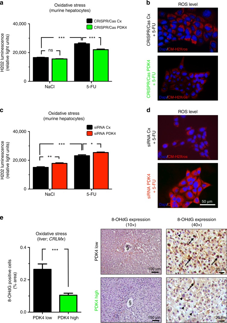
Fig. 5.
Up-regulation of PDK4 attenuated chemotherapy-induced oxidative stress. a–d Hydrogen peroxide (H2O2) luminescence assays (a, c) and representative fluorescence labelling for CM-H2Xros (red) and DAPI (cell nuclei, blue) (b, d) to indicate oxidative stress in murine hepatocytes (AML12) exposed to 5-FU (500 µM) or vehicle control (NaCl). Note that 5-FU-induced oxidative stress is significantly attenuated by up-regulation of PDK4 gene function (CRISPR/Cas PDK4) compared to control (CRISPR/Cas Cx) (a, b), but aggravated by siRNA-mediated knockdown of PDK4 (siRNA PDK4) compared to control (siRNA Cx) (c, d). Graphs (a, c) represent pooled data from three independent experiments. *P < 0.05; **P < 0.01; ***P < 0.001; n = 3. e Representative 8-OHdG immunostainings (right) and histomorphometric quantification (left) of liver tissue from CRLM patients (from CRLMx cohort), indicating significantly attenuated oxidative stress in patients with high hepatic PDK4 expression (green bar and bottom panels) compared to those displaying low hepatic PDK4 expression (black bar and upper panels). Note reduced nuclear abundance (arrowheads in right panels) of 8-OHdG adducts (arrows in right panels) in patients with high hepatic PDK4 expression. n = 8 per group; ***P < 0.001. The sectional symbol indicates central vein.

