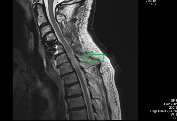
Fig. 4.
Postoperative sagittal T2 magnetic resonance imaging image demonstrates a wide posterior decompression of the cervical spinal cord. A huge intramedullary hypertense area, exactly below the C6 vertebrae (green arrow) after the second surgery to our department approximately 24–28 h after the first surgery

