Fig. 6.
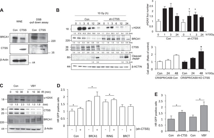
CTSS decreases DNA damage responses. a Control or WT-CTSS was transiently transfected into MCF7 cells. The levels of BRCA1 and CTSS in the dsDNA pull-down lysates, as well as in complete whole nuclear extracts, were analyzed. b MCF7 cells were irradiated with 10 Gy IR after transfection with control or sh-CTSS, and Western blotting was performed at indicated time points. Protein levels were quantified using Image J software, and data are expressed as the fold change relative to the negative control (left). Immunofluorescence analysis for γH2AX foci were performed after 10 Gy IR (right upper). Apoptotic cells after 10 Gy IR were evaluated by PI staining in CRISPR-Cas9 CTSS KO MCF7 cell lines. *p < 0.01 vs. corresponding control cells (ANOVA) (right bottom). c After 10 Gy radiation, Western blotting was performed at indicated time points with or without treatment of CTSS-specific inhibitor VBY-036 (10 μM). d The ratio of GFP + cells transfected with sh-CTSS, BRCA1, ΔBRCT, or ΔRING expression in MCF7 cells that stably expressed DR-GFP were analyzed by FACS. e GFP + cells after treatment of VBY-036 (10 μM) or sh-CTSS in MCF7 cells that stably expressed DR-GFP were analyzed by FACS (mean ± SD from 3 different experiments). *p < 0.05 (ANOVA)

