Abstract
Background
Rapamycin (alternatively known as sirolimus) is a macrolide immunosuppressant commonly used for organ transplantation. It acts both on lymphocytes through the mechanistic target of rapamycin (mTOR) pathway to reduce their sensitivity to interleukin-2 (IL-2) and, importantly, also has anti-fibrotic properties by acting on myofibroblasts. The latter have been implicated in the pathogenesis of thyroid eye disease (TED).
Aim
To describe successful treatment and reversal of extraocular muscle fibrosis in TED with sirolimus.
Methods
Case report and literature review with clinic-pathological correlation.
Results
A patient with Graves’ orbitopathy (GO) developed significant ocular motility restriction, which was unresponsive to steroids and conventional immunosuppression. Unlike these prior treatments, rapamycin therapy improved the diplopia and fields of binocular single vision over a period of months. There were no adverse effects directly attributable to the treatment.
Conclusion
With its low renal toxicity and ability to specifically target the underlying fibrotic pathways in GO, rapamycin may prove a useful adjunct to standard immunosuppressive regimes. We encourage further reporting of case series or the instigation of controlled trial.
Introduction
Rapamycin (sirolimus) is a potent immunosuppressive agent with anti-proliferative and anti-fibrotic actions. Unlike other immune suppressants, it is associated with minimal renal toxicity and side effects are uncommon at low doses, wherefore it has been widely used long term for the prevention of transplant rejection.
Sirolimus acts by binding to FK-binding-protein-12 to inhibit a subunit protein of the mechanistic target of rapamycin called complex 1 (mTORC1), which is essential for T cell activation and which has also been implicated in adipogenesis [1]. In vitro studies have additionally demonstrated anti-fibroblast effects of rapamycin, including reduction in fibroblast migration and reduced transition to myofibroblasts [2], proliferation, collagen expression, interleukin-16 (IL-16) production [3] and cellular metabolism [4]. In vivo, murine experiments indicate reduced wound repair and fibrosis with immune suppression [5] as well as reduced fibrosis in a variety of organ systems including kidney [6] and heart [7] with rapamycin treatment.
Overall, the evidence suggests that mTOR plays a significant role in the control of abnormal fibroblast proliferation, differentiation and extrusion of stromal contents including collagen [2, 4]. Because of its attenuation of adipogenesis and effect on fibroblast proliferation and fibrosis—processes all implicated in the pathogenesis of Graves’ orbitopathy (GO) [1]—we sought to determine whether rapamycin could be a potential adjunct in the treatment of thyroid eye disease (TED). Tantalisingly, it has been shown that downstream signalling from the insulin-like growth factor-1 (IGF-1) receptor is also blocked by rapamycin [8]—and insulin-like growth factor receptor (IGFR) too is strongly implicated in the pathogenesis of TED [9].
To date, only a single patient with TED has been reported to have had treatment with sirolimus—a case that was described over a decade ago; [10] the authors found rapamycin to be effective for the treatment of dysthyroid compressive optic neuropathy, which had proven refractory to orbital decompression surgery and worsened with steroid treatment [10]. However, a reduction in fibrosis, such as increased excursions of fibrosed extraocular muscles, has not been previously demonstrated with rapamycin.
Case
A 43-year-old male smoker (30 cigarettes a day) but with no other past medical or family history, presented with a 10-month history of Graves’ thyrotoxicosis and 4-month history of painless ‘white-eyed’ diplopia. Examination showed significant ocular motility restriction in all positions of gaze, but visual acuities were unaffected, and apart from mild conjunctival injection, there were no other signs of active inflammation. There was only minimal right proptosis of <2 mm, and no evidence of exposure keratopathy or optic neuropathy (no relative afferent pupillary defect, normal colour vision, full automated perimetry). Magnetic resonance imaging confirmed prominence of all the extraocular muscles with orbital apex crowding and increased signal on T2-weighted images particularly in the right inferior rectus.
His endocrine disease had been treated with carbimazole titration by the referring unit where, given the marked ocular motility restriction, he was also started on oral prednisolone 80 mg/day tapering to 30 mg over 2 months. The ocular motility worsened with steroid therapy and this active smoker was therefore referred to our tertiary service for management.
The patient was borderline hypothyroid [thyroid-stimulating hormone (TSH) 11.2 (relative risk (RR) 0.35–5.50 mU/L), T4 10.3 (10.0–19.8 pmol/L), thyroid peroxidase antibody (TPO) 18 (0–60 IU/mL), anti-TSH receptor antibody (TRAb) 0.9 (0.0–1.0 IU/L)] and was switched to a block and replace regimen by the addition of levothyroxine. There was no evidence of dysthyroid optic neuropathy (DON) but all binocular single vision (BSV) was now lost, with complete loss of upgaze on the right. With the minimal signs of inflammation and low TRAb, magnetic resonance imaging (MRI) T2-weighted imaging was arranged to confirm post-inflammatory fibrosis as the likely cause of the worsening motility restriction.
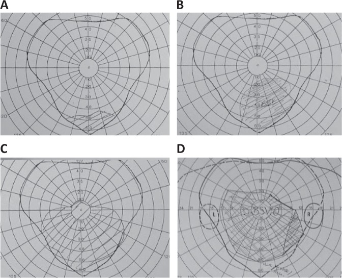
Given the degree of fibrosis, sirolimus 6 mg then 4 mg daily was started in preference to cyclosporine, which is our usual second-line agent [11]. Trough levels of 6–12 ng/mL were rapidly achieved and within 2 weeks there was a small inferior island of BSV on adoption of a chin up head posture, although there was no subjective improvement (see Fig. 1a). The oral prednisolone was tapered and blood tests showed no adverse impact of treatment on kidneys, liver or other organs. Further improvement in the fields of BSV was observed over the 15 months to reach a stable point (see Fig. 1b, c). Steroids and sirolimus were then both tapered over several months. The patient remained inactive and maintained good BSV (see Fig. 1d) and had addition of a prism to eliminate head posture. He was then discharged back to his referring unit.
Fig. 1.
Plot of field of binocular single vision (BSV) testing performed 2 weeks after starting sirolimus, on adopting an abnormal head posture (a). A small island of BSV subtending 4 quadrilaterals has appeared inferiorly indicated by the shaded area. b shows an increase in the area of BSV to 17 quadrilaterals, 8 months after starting sirolimus and performed with a reduced chin up head posture. c Fifteen months after the start of treatment, the BSV has increased to 46 quadrilaterals and the test was performed without a compensating head posture. d The effect persists even after cessation of treatment, seen here 3 years later with a BSV subtending 63 quadrilaterals
Discussion
Rapamycin was the first inhibitor of mTORC1 to be discovered and the drug itself, sirolimus, was approved by the United States Food and Drug Administration (FDA) in 1999 for the prevention of transplant rejection [12], and thereafter, from 2007 onwards, the treatment of certain cancers including renal cell carcinoma. It was at this time that our patient presented with significant GO with marked restriction of ocular motility. Clinical assessment and MRI signal intensity on T2 were not suggestive of marked activity, and the decision was made to use sirolimus to limit further fibrosis in the extraocular muscles, whilst also allowing withdrawal of steroid.
Orbital fibroblasts are amongst the main effector cells in the pathogenesis of thyroid eye disease, yet few therapies are known to target them preferentially. Here we report results to suggest that sirolimus can be a helpful adjunct to prednisolone-based immunosuppression, with the possibility of improving ocular motility in the post-inflammatory, fibrotic, stage of GO. Conventional immunosuppressive agents can have significant side effects or fail to be efficacious for a subset of patients with TED. So targeting the tissue remodelling which is seen in GO may prove an alternative attractive avenue.
This has been attempted once previously: in 2007, Chang et al. [10] reported a single patient with DON who was treated successfully with sirolimus. However the results were confounded by the patients’ prior thyroid ablation, decompression surgery, steroid therapy and regular use of rosiglitazone and atorvastatin. In contrast, our patient had less active disease, no other medical history and had worsened on steroid therapy alone. Furthermore, whilst the orbitopathy treated by Chang et al. [10] was characterised by adipogenesis and compartment expansion, our patient was affected principally by fibrotic changes in the muscle without volume and compartment problems. Despite the different phenotype, sirolimus proved effective also for our patient. Improvement in BSV was quantified by counting the shaded quadrilaterals, which make up the grid in the fashion described by Fitzsimons and White [13]. Within 2 weeks of starting sirolimus, the number of quadrilaterals had increased from 0 to 4, then 17, 46 and 63 at the last visit.
At higher doses, sirolimus has been reported to have a number of side effects, including hypertriglyceridaemia, hypertension, hypercholesterolaemia, increased creatinine, thrombocytopenia and diabetes. In our patient, glycosuria was noted prior to the introduction of sirolimus, but with careful monitoring of trough levels, glycaemia and haemoglobin A1c, treatment was not required. As with other immunosuppressant regimes, full blood count, urea and electrolytes, lipid profile, trough levels, assessment for impaired glucose tolerance and hypertension are important prior to initiating therapy. Fortuitously, however, treatment is effective for TED at lower doses than those used for transplantation [10].
Successful treatment with sirolimus also sheds light on the molecular pathogenesis of thyroid eye disease. The activity of the mTORC1 complex has been known to be regulated by rapamycin, insulin, IGFR, basal metabolic rate, energy availability and oxidative stress, suggesting a possible mechanistic link between the increased incidence of TED in those with poorly controlled thyroid function or who smoke.
Significant progress has been made in the understanding of the molecular pathways involved. Unlike other immune suppressants, rapamycin acts to block both signal transduction and cell cycle progression [10], the implication being that it can have an additive effect with other therapies. Recent pre-clinical studies also supports this; targeting the phosphatidylinositol 3-kinase (PI3K) and mTORC1 pathways concurrently might be suitable for non-immunosuppressive therapy of GO by limiting adipogenesis, inflammation and hyaluronic acid accumulation [14]. mTORC1 is essential for adipogenesis, whereas PI3K signalling specifically regulates haemagglutinin (HA) production via HA synthase 2 (HAS2), the major contributor of HA in the orbit. Zhang et al. [14] found that targeting both the PI3K and mTORC1 pathways was possible with a single agent—trifluoperazine hydrochloride—in vitro. This may represent a future alternative to sirolimus therapy alone.
The need for improved means by which to assess clinical activity in TED is also highlighted by this case. We used MRI T2-relaxation times with fat fraction mapping [15] in combination with circulating TRAb levels [16] to determine that the disease in our patient was still active despite the patient’s clinical activity score [17] being low. With his ‘white eye’, it would have been relatively easy to assume disease inactivity. However, initial MRI confirmed significant disease with activity, and this prompted the decision to commence immunosuppression. An initial TRAb was not available from the referring unit, but on review in our centre, a normal TRAb and improvement in MR T2 signal, despite worsening motility restriction, was able to reassure that activity was improving on steroid treatment, but that a second-line agent that could limit fibrosis and allow steroid withdrawal would be useful.
Further studies are required before rapamycin can be established as a suitable agent for reduction of fibrosis and adipogeneis in GO, but this report suggests that it could be a potentially useful agent, and adds support for the need to explore treatments that target the mTORC1 pathway.
Acknowledgements
The authors would like to thank Dr. Paul Meyer.
Author contributions
All authors have contributed to the data collection and writing.
Conflict of interest
All authors are doctors who manage patients with thyroid eye disease. The authors declare that they have no conflict of interest.
Ethics statement
As an anonymised retrospective report no institutional review board authorisation was necessary.
Guarantor
Dr. Murthy serves as guarantor of this work. It is an honest, accurate and transparent account of the study being reported; no important aspects of the study have been omitted.
Footnotes
License: The corresponding author has the right to grant on behalf of all authors and does grant on behalf of all authors, an exclusive licence on a worldwide basis to permit this article to be published.
Publisher’s note: Springer Nature remains neutral with regard to jurisdictional claims in published maps and institutional affiliations.
References
- 1.Zhang L, Grennan-Jones F, Lane C, Rees DA, Dayan CM, Ludgate M. Adipose tissue depot-specific differences in the regulation of hyaluronan production of relevance to Graves’ orbitopathy. J Clin Endocrinol Metab. 2012;97:653–62. doi: 10.1210/jc.2011-1299. [DOI] [PubMed] [Google Scholar]
- 2.Molina-Molina M, Machahua-Huamani C, Vicens-Zygmunt V, Llatjós R, Escobar I, Sala-Llinas E, et al. Anti-fibrotic effects of pirfenidone and rapamycin in primary IPF fibroblasts and human alveolar epithelial cells. BMC Pulm Med. 2018;18:63. doi: 10.1186/s12890-018-0626-4. [DOI] [PMC free article] [PubMed] [Google Scholar]
- 3.Pritchard J, Horst N, Cruikshank W, Smith TJ. Igs from patients with Graves’ disease induce the expression of T cell chemoattractants in their fibroblasts. J Immunol. 2002;168:942–50. doi: 10.4049/jimmunol.168.2.942. [DOI] [PubMed] [Google Scholar]
- 4.Hillel AT, Gelbard A. Unleashing rapamycin in fibrosis. Oncotarget. 2015;6:15722–3. doi: 10.18632/oncotarget.4652. [DOI] [PMC free article] [PubMed] [Google Scholar]
- 5.Ghosh A, Malaisrie N, Leahy KP, Singhal S, Einhorn E, Howlett P, et al. Cellular adaptive inflammation mediates airway granulation in a murine model of subglottic stenosis. Otolaryngol Head Neck Surg. 2011;144:927–33. doi: 10.1177/0194599810397750. [DOI] [PubMed] [Google Scholar]
- 6.Chen G, Chen H, Wang C, Peng Y, Sun L, Liu H, et al. Rapamycin ameliorates kidney fibrosis by inhibiting the activation of mTOR signaling in interstitial macrophages and myofibroblasts. PLoS ONE. 2012;7:e33626. doi: 10.1371/journal.pone.0033626. [DOI] [PMC free article] [PubMed] [Google Scholar]
- 7.Yu SY, Liu L, Li P, Li J. Rapamycin inhibits the mTOR/p70S6K pathway and attenuates cardiac fibrosis in adriamycin-induced dilated cardiomyopathy. Thorac Cardiovasc Surg. 2013;61:223–8. doi: 10.1007/s11748-012-0131-2. [DOI] [PubMed] [Google Scholar]
- 8.Krassas GE, Gogakos A. Thyroid-associated ophthalmopathy in juvenile Graves’ disease—clinical, endocrine and therapeutic aspects. J Pediatr Endocrinol Metab. 2006;19:1193–206. doi: 10.1515/JPEM.2006.19.10.1193. [DOI] [PubMed] [Google Scholar]
- 9.Smith TJ, Kahaly GJ, Ezra DG, Fleming JC, Dailey RA, Tang RA, et al. Teprotumumab for thyroid-associated ophthalmopathy. N Engl J Med. 2017;376:1748–61. doi: 10.1056/NEJMoa1614949. [DOI] [PMC free article] [PubMed] [Google Scholar]
- 10.Chang S, Perry JD, Kosmorsky GS, Braun WE. Rapamycin for treatment of refractory dysthyroid compressive optic neuropathy. Ophthalmic Plast Reconstr Surg. 2007;23:225–6. doi: 10.1097/IOP.0b013e3180500d57. [DOI] [PubMed] [Google Scholar]
- 11.Roos Jonathan C. P., Murthy Rachna. Comment on: A British Ophthalmic Surveillance Unit (BOSU) study into dysthyroid optic neuropathy in the United Kingdom. Eye. 2018;33(2):327–342. doi: 10.1038/s41433-018-0303-0. [DOI] [PMC free article] [PubMed] [Google Scholar]
- 12.Nashan B, Citterio F. Wound healing complications and the use of mammalian target of rapamycin inhibitors in kidney transplantation: a critical review of the literature. Transplantation. 2012;94:547–61. doi: 10.1097/TP.0b013e3182551021. [DOI] [PubMed] [Google Scholar]
- 13.Fitzsimons R, White J. Functional scoring of the field of binocular single vision. Ophthalmology. 1990;97:33–5. doi: 10.1016/S0161-6420(90)32631-3. [DOI] [PubMed] [Google Scholar]
- 14.Zhang L, Ji QH, Ruge F, Lane C, Morris D, Tee AR, et al. Reversal of pathological features of graves’ orbitopathy by activation of forkhead transcription factors, FOXOs. J Clin Endocrinol Metab. 2016;101:114–22. doi: 10.1210/jc.2015-2932. [DOI] [PubMed] [Google Scholar]
- 15.Das Tilak, Roos Jonathan C. P., Patterson Andrew J., Graves Martin J., Murthy Rachna. T2-relaxation mapping and fat fraction assessment to objectively quantify clinical activity in thyroid eye disease: an initial feasibility study. Eye. 2018;33(2):235–243. doi: 10.1038/s41433-018-0304-z. [DOI] [PMC free article] [PubMed] [Google Scholar]
- 16.Roos Jonathan C. P., Paulpandian Vignesh, Murthy Rachna. Serial TSH-receptor antibody levels to guide the management of thyroid eye disease: the impact of smoking, immunosuppression, radio-iodine, and thyroidectomy. Eye. 2018;33(2):212–217. doi: 10.1038/s41433-018-0242-9. [DOI] [PMC free article] [PubMed] [Google Scholar]
- 17.Mourits MP, Koornneef L, Wiersinga WM, Prummel MF, Berghout A, van der Gaag R. Clinical criteria for the assessment of disease activity in Graves’ ophthalmopathy: a novel approach. Br J Ophthalmol. 1989;73:639–44. doi: 10.1136/bjo.73.8.639. [DOI] [PMC free article] [PubMed] [Google Scholar]


