
Fig. 5.
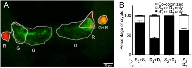
Strain localization in the crypts of the light organ. Squids were inoculated with one of four different strain combinations: (i) two S strains, (ii) two D strains, or (iii) an S strain, with a D strain added either simultaneously, or (iv) after a 3 h delay. (a) After 48 h, squids were fixed, and their light organs observed under a confocal microscope to determine the percentage of their 6 colonized crypts (gray outline) that were singly colonized by a GFP- (G) or an RFP- (R) labeled strain or were co-colonized by both of the inoculated strains (GR). (b) For each treatment, 53 or 54 animals were analyzed in four replicates, corresponding to between 187 and 217 colonized crypts observed per treatment. The bar graph represents the mean percentage of each outcome, and the error bars the 95% confidence interval. The crypts analyzed were either co-colonized (gray) by both of the inoculating strains, or were only singly colonized by one of the pair of strains (white) or the other (black)