Fig. 5.
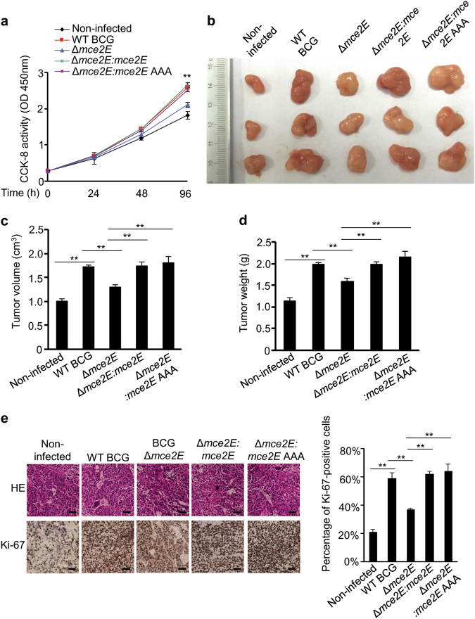
Mtb Mce2E promotes A549 cell proliferation in a D motif-independent manner. a CCK-8 assay of A549 cells. Cells were infected with WT BCG, BCG Δmce2E, BCG Δmce2E:mce2E, or BCG Δmce2E:mce2E AAA at a MOI of 10 for 0–4 days, and then, the OD450nm value was measured and cell numbers were calculated. Non-infected A549 cells were used as a control. b Photograph of tumors. Tumors were derived from non-infected A549 cells or cells infected as in a in BALB/c nude mice. c Volume of tumors in nude mice. Tumor cells were injected subcutaneously into the right armpit of nude mice, the short and long diameters of the tumors were measured and the tumor volumes (cm3) were calculated at 14 days after injection. d Weight of tumors in nude mice. e Representative H&E staining histopathologic images of tumor tissues from mice (upper panels) and immunohistochemical analysis of Ki-67 (lower panels). Scale bars, 200 μm. Right, percentage of Ki-67-positive cells in tumors. Approximately 200 cells were counted. *P < 0.05 and **P < 0.01 (unpaired two-tailed Student’s t test). Data are representative of experiments with three independent biological replicates (mean and s.e.m. of n = 6 mice per group)

