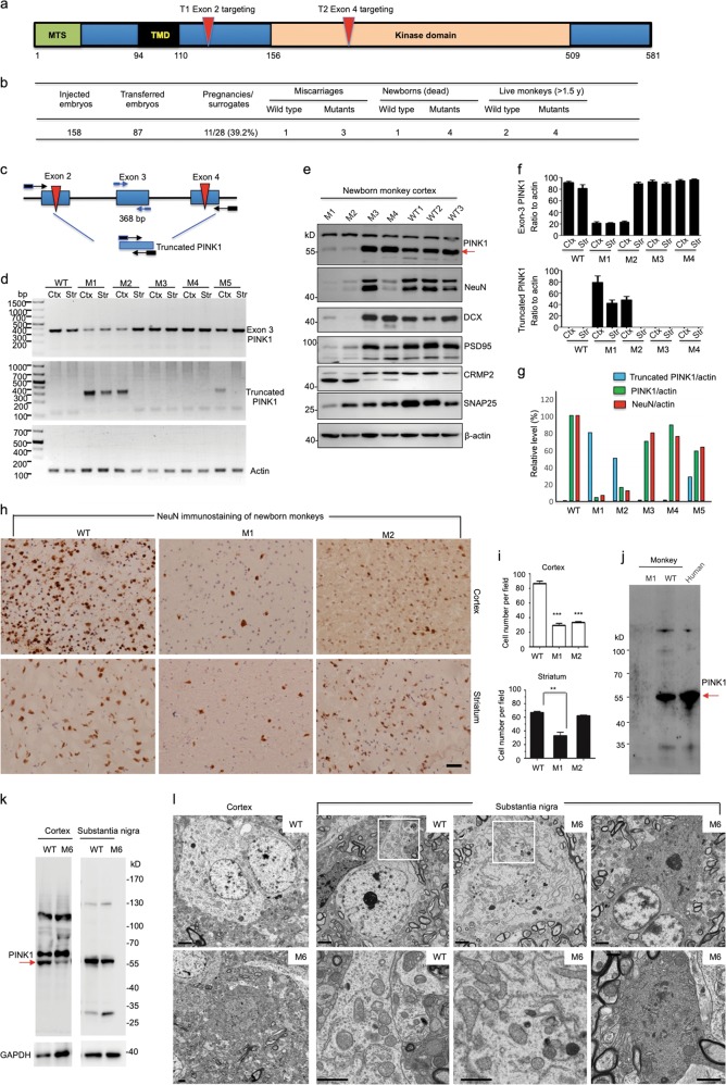
Fig. 1.
a PINK1 protein domains and targeted regions. MTS (mitochondrial targeting sequence), TMD (transmembrane domain), exon 2 (T2) and exon 4 (T4) targeting regions are indicated. b Summary of embryo injection, transfer, pregnancy, and newborn monkeys. c Diagram of PCR primers designed to determine the PINK1 large deletion. d Large PINK1 deletion in the cortex and striatum of M1, M2, M3, M4, and M5 monkeys. Exon 3 PINK1 represents the remaining intact PINK1, whereas truncated PINK1 is generated by large deletion. e Western blot analysis of the brain cortical tissues of four PINK1 mutant monkeys and three newborn wild-type (WT) monkeys. The tissues were probed with antibodies to PINK1, neuronal proteins (NeuN, PSD95, CRMP2, and SNAP25), β-actin, and doublecortin (DCX). f Quantitative analysis of the ratio of exon 3 PINK1 or truncated PINK1 product to actin revealed that the cortex (Ctx) and striatum (Str) of M1 and the cortex of M2 had an extensive large deletion. The results were obtained from three PCR experiments. g Inverse correlation of the rate of the large PINK1 deletion (truncated PINK1/actin) with the relative levels of PINK1 (PINK1/actin) or NeuN (NeuN/actin) revealed by western blotting (n = 3 independent experiments via analysis of the cortical tissues in three WT and four mutant monkeys). h Representative immunostaining micrographs show loss of NeuN-positive neurons in the cortex of PINK1 mutant monkeys (M1 and M2) compared with WT controls. Scale bar, 30 μm. i Quantitative assessment of NeuN-positive neurons in the cortex and striatum of newborn PINK1 mutant monkeys (M1 and M2) and two WT control monkeys. **P < 0.05, ***P < 0.01. j Western blotting of lysates from WT or M1 monkey brain cortex and the human brain hippocampus. Red arrow indicates PINK1. k Western blot analysis of cortex and substantia nigra lysates of M6 monkey and age-matched 3-year-old WT monkey. Red arrow indicates PINK1. l Electron microscopy revealed degenerated cells in the cortex and substantia nigra of the M6 monkey. Degenerated neurons show electron-dense cytoplasm with no clear organelles and no identifiable nuclear membrane, and dark neurons are more obvious in the M6 substantia nigra. The enlarged micrographs beneath the corresponding WT and M6 images are from their boxed areas and show mitochondrial morphology. Scale bars, 2 μm

