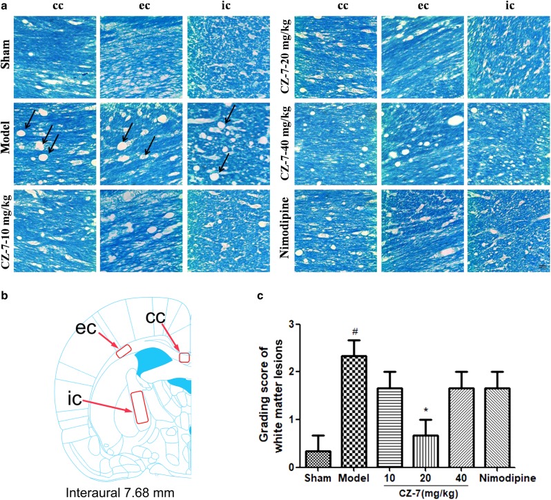
Fig. 6.
Effects of CZ-7 on chronic cerebral hypoperfusion-induced white matter lesions. a Representative photographs of tissue sections stained with Klüver–Barrera in the myelinated fibers of the corpus callosum (cc), external callosum (ec), and internal capsule (ic). Arrows indicate the formation of marked vacuoles and the disappearance of myelinated fibers. b A schematic elucidation of the rat cc, ec, and ic. Red boxes represent the observed areas of all brain slices in a. c Grading score of white matter lesion. Values are expressed as mean ± SEM. n = 3. #P < 0.05 vs. Sham group, *P < 0.05 vs. Model group. For grading score of white matter lesion, one-way ANOVA, F (5, 17) = 4.967, P = 0.0107

