Fig. 2.
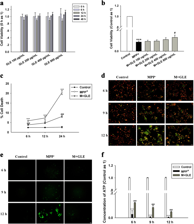
GLE treatment prevented cellular and mitochondrial damage in neuro-2a cells following MPP+ injury. Neuro-2a cells were exposed to MPP+ (1 mM, at the indicated time point) with or without GLE. a Cytotoxic effect of GLE at varying concentrations and incubation times on neuro-2a cells. b Effect of GLE on the MPP+-induced reductions in neuro-2a cell viability. Multiple concentrations of GLE were used to determine the concentration that was non-toxic to cells but caused the most significant increase in cell viability in the case of MPP+ injury; 800 μg/mL was the dose of GLE selected for subsequent trials. c Percentage of dead cell was determined by the trypan blue membrane permeability assay as described in the Materials and Methods. d Representative image of mitochondria membrane potential (MMP) staining in each group. MMP was assessed by JC-1 dye. e Representative fluorescence microscopy image of neuro-2a cells stained with ROS-detecting probes (green). f Concentrations of intracellular ATP using an ATP Determination Kit. Data are expressed as the mean ± SEM (at least three independent experiments were performed); ***P < 0.001 vs. the control group; #P < 0.05, ###P < 0.001 vs. the MPP+ group

