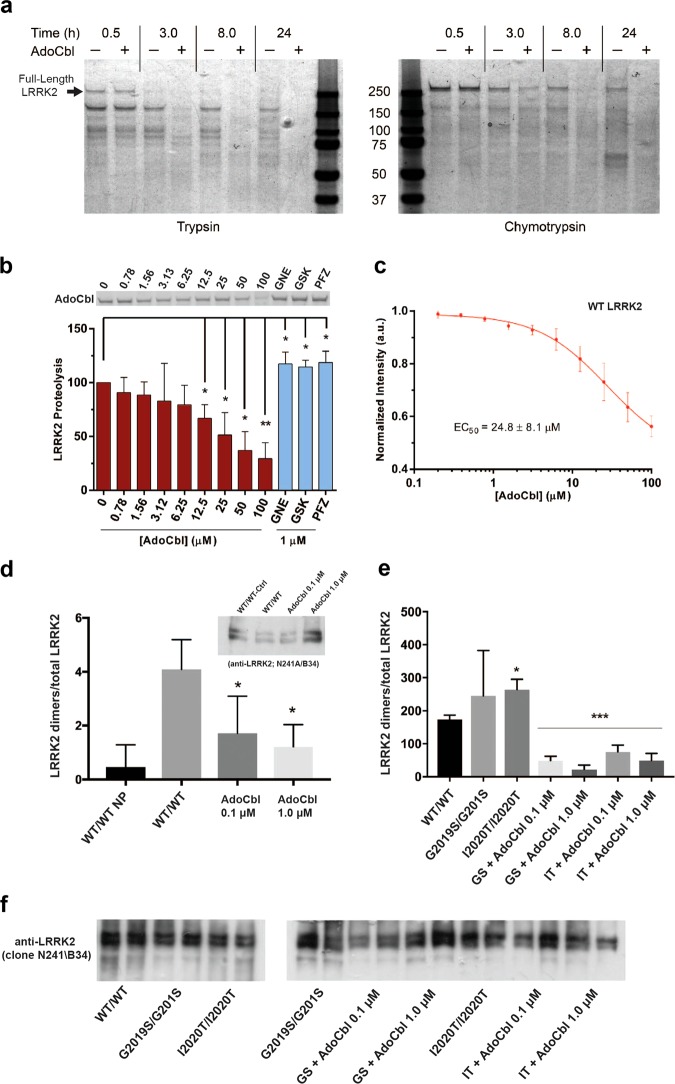
Fig. 4.
AdoCbl causes a LRRK2 conformational change and destabilizes LRRK2 dimers. a Coomassie stained SDS-PAGE showing limited proteolysis analysis using a 10:1 molar ratio of LRRK2: Trypsin (left panel) and LRRK2: Chymotrypsin (right panel). Proteolysis was performed at 30 °C with or without 50 µM AdoCbl and reactions were quenched at the indicated times by the addition of sample loading buffer. The observed data were consistent across three biological replicates. b Limited proteolysis of LRRK2-WT by trypsin in the presence of increasing concentrations of AdoCbl, or 1 µM LRRK2 kinase inhibitor. Proteolysis was performed for 90 min at 30 °C. Shown is a representative SDS-PAGE of full-length LRRK2, in which bands were quantified and values were normalized to LRRK2 proteolysis without AdoCbl. c The peak intrinsic fluorescence of LRRK2 (339 nm) was measured as a function of AdoCbl. Strep-tagged LRRK2 was incubated with indicated concentrations of AdoCbl for 30 min prior to fluorescence measurements. Significance was measured by one-way ANOVA. *p ≤ 0.05, **p ≤ 0.005. d HEK293T cells co-expressing BirA-WT (biotin ligase) and AP-WT LRRK2 (acceptor peptide) were lysed following a biotin pulse to label dimeric LRRK2, and extracts bound to streptavidin-coated ELISA plates. LRRK2 was detected using anti-LRRK2 conjugated to HRP (clone N241A/B34) and expressed as a ratio of total LRRK2 levels detected by ELISA in parallel plates coated with total LRRK2 antibodies (clone c41-2). In the plot, “WT/WT NP” refers to cells expressing WT LRRK2 dimers that were harvested without receiving a biotin pulse (“no pulse”). AdoCbl significantly reduced levels of dimeric WT-LRRK2. Sub-panel shows representative immunoblot of parallel extracts detected with anti-LRRK2 (clone N241A/B34). BirA-LRRK2 represents the top band, and AP-LRRK2 the bottom band. e HEK293T cells expressing BirA- G2019S or I2020T mutant LRRK2 together with AP-G2019S or I2020T LRRK2, and dimeric LRRK2 quantified by ELISA. Treatment with AdoCbl significantly reduces dimeric mutant LRRK2. *p < 0.05 compared to WT/WT-LRRK2; ***p < 0.001 compared to G2019S-LRRK2 dimers or I2020T-LRRK2 dimers alone. f Representative immunoblot of parallel extracts detected with anti-LRRK2 (clone N241A/B34). BirA-LRRK2 represents the top band, and AP-LRRK2 the bottom band

