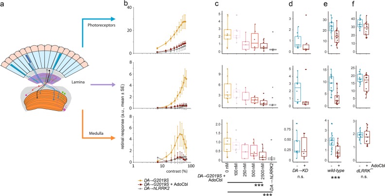
Fig. 6.
AdoCbl rescues deficits in Drosophila visual physiology induced by the dopaminergic expression of human LRRK2-G2019S. a Outline of the retinal neural network of Drosophila, with three main neuronal layers: photoreceptors, lamina neurons, and medulla neurons (Modified after Afsari et al.31) b Contrast response functions (CRFs) for the photoreceptors, lamina neurons and medulla neurons show that the dopaminergic expression of hLRRK2-G2019S (DA →G2019S) flies have a much stronger response than either the DA→hLRRK2 or the DA→G2019S which have been fed 2.5 μM AdoCbl. c Dose-response curve for the effect of AdoCbl on the DA→G2019S flies, shows a 50% reduction in phenotypes by 250–500 nM AdoCbl, with almost complete rescue by 2.5 μM AdoCbl. d There is no effect of 2.5 μM AdoCbl on flies with dopaminergic expression of kinase-dead hLRRK2-G2019S-K1906M (DA →KD). e The visual response of flies with wild-type dLRRK2 is reduced by 2.5 μM AdoCbl. f Applying 2.5 μM AdoCbl to dLRRK¯ transheterozygote flies (in which the Drosophila LRRK2 homolog has been knocked out) has no statistically significant effect. Data represent the mean (±s.e.) and the dots represent the number of flies tested. In c, statistical analysis from Tukey Post-hoc tests on the first principal component of a PCA, which accounted for 88% of the variance (Supplementary information, Fig. S9). d–f analysis by MANOVA. n.s. not significant; ***p < 0.001). Boxes correspond to the median ± quartiles. Dots indicate data from individual flies. dLRRK¯ genotype: dLRRKe03680/dLRRKex1; wild-type genotype: wa/w1118

