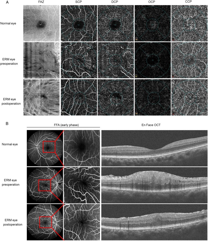
Fig. 2.
Pre-operative and post-operative OCTA and FFA analyses of an ERM patient. a Representative images were shown from the normal contralateral eye and the affected eye in a patient with ERM before and after surgery using OCTA. The overall OCTA morphology of the fovea was presented as follows: SCP, DCP, OCP, and OCP. Post-operative evaluation was performed at 6 months after therapy. b Representative images of FFA and En Face OCT images were shown in a case of pre-operative and post-operative ERM eye and normal fellow eye. FFA images were collected at the early phase post dye injection and the macula was magnified to show vascular distortion as in the figure. No leakage was found in pre-operative and post-operative eyes at the early stage of FFA. OCTA optical coherence tomography angiography, SCP superficial capillary plexus, DCP deep capillary plexus, OCP outer retinal capillary plexus, CCP choroidal capillary plexus

