Fig. 5.
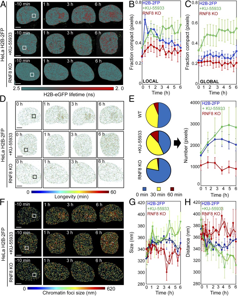
ATM and RNF8 regulate chromatin architecture in the DDR. (A) FLIM-FRET maps of HeLaH2B-2FP, HeLaH2B-2FP treated with KU-55933, or HeLaH2B-2FP RNF8 KO cells 10 min before and 1, 3, and 6 h after microirradiation. (B and C) Fraction of compacted chromatin pixels within (B) and outside (C) of the NIR irradiation site in HeLaH2B-2FP (blue curve; n = 10), KU-55933-treated (green curve; n = 6), or RNF8 KO cells (red curve; n = 3; mean ± SEM). (D) Longevity maps of compact chromatin foci from the cells in A. (E) Quantification of compact chromatin foci stability (Left) throughout the DDR (Right) in NIR laser-treated HeLaH2B-2FP, KU-55933–treated HeLaH2B-2FP, and HeLaH2B-2FP RNF8 KO cells (n as indicated above, mean ± SEM). (F) Chromatin compaction size map derived by PLICS from the cells shown in A. (G and H) Change in the mean size of (G) and distance between (H) compacted chromatin foci during the DDR in NIR laser-treated HeLaH2B-2FP, KU-55933–treated HeLaH2B-2FP, and HeLaH2B-2FP RNF8 KO cells (n as indicated above, mean ± SEM). (Scale bars, 5 μm.)

