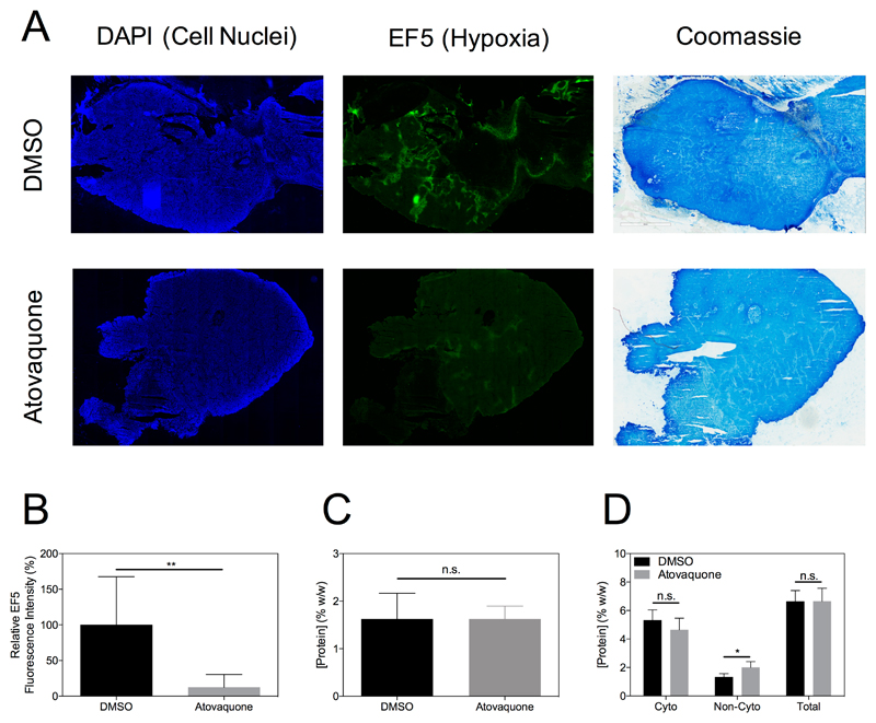
Figure 6.
Representative tissue sections from tumours in the DMSO or Atovaquone treatment groups stained for DAPI, EF5 and Coomassie show reduction in tumour hypoxia due to Atovaquone with no concomitant alteration of protein concentration (A). The reduction in tumour hypoxia is evident as a significant reduction in EF5 fluorescence intensity across all animals (B, P < 0.01, unpaired t-test). Cytoplasmic protein concentration as measured by Coomassie staining (C) or BCA assay (D) was not significantly different between Atovaquone and DMSO groups (P > 0.05, unpaired t-test).

