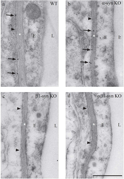
Figure 4: Electron micrographs showing immunogold labeling for Kir4.1 in perivascular endfoot domain of Müller cell.

Immunogold labeling of Kir4.1 can be seen along the length of the perivascular endfoot process of Müller cells in WT mice (a). In mice lacking α1-syntrophin, the labeling is retained (b). Absence of perivascular Kir4.1 labeling is seen in β1-syn KO (c) and in αβ1-syn KO retinae (d). The arrowheads indicate the Müller cell endfoot domain facing the blood vessel. Scale bar=500nm; L, Lumen; E, Endothelium; *=Basement membrane.
