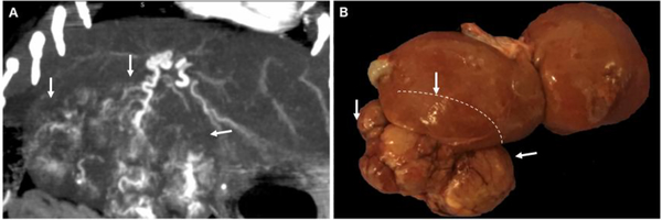
Figure 1:
Spontaneous HCC developing in chronically hepatitis infected woodchucks. (A) Contrast-enhanced CT scan shows large heterogenous tumor with robust arterial blood supply (B). Gross pathology of liver and tumor (tumor edges demarcated by white arrows, margins of the tumor behind the liver demarcated by dashed line).

