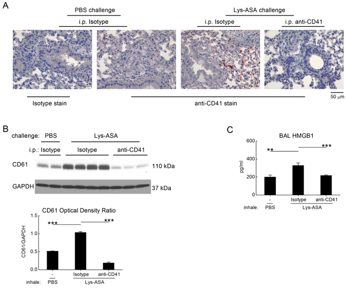
Figure 4. Recruited platelets account for the Lys-ASA-induced increment in HMGB1.
Lungs were collected from PBS- or Lys-ASA-challenged Ptges−/− mice treated with a platelet-depleting anti-CD41 Ab or isotype control. A. Lung sections were stained with anti-CD41 mAb or an isotype control. Infiltrating platelets are identified by the red staining. B. Western blotting of lung lysates from Ptges−/− mice of the indicated treatment groups for CD61 as a surrogate marker for recuited platelets. A blot from a representative individual experiment (top) and quantitative densitometry from two separate experiments (bottom) are shown. C. BAL fluid levels of HMGB1 from mice challenged with Lys-ASA or PBS after treatment with anti-CD41 or isotype. Results in A are from single mice representative of at least 5/group in three different experiments. Results in C are mean ± SEM from at least 10 mice/group from two separate experiments.

