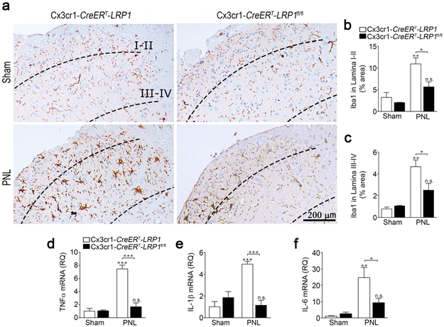
FIGURE 6.
LRP1 deletion in microglia in adult mice attenuates microglial activation in the SDH following PNL. (a) TAM-treated Cx3cr1-CreERT-LRP1fl/fl (LPR1fl/fl) mice and Cx3cr1-CreERT-LRP1 (LRP1) mice were subjected to PNL or sham-operation. SDH tissue was isolated 3 days later. Representative IHC images for Iba1 are shown. (b, c) Densitometry analysis was performed to assess the percentage of tissue area occupied by Iba1 immunostaining in Laminae I-II and in Laminae III-IV (mean ± s.e.m.; n = 3 for sham-operated animals and n = 4 for animals subjected to PNL;*p < 0.05, **p < 0.01, n.s. not significant, the signs directly over the bars compare PNL with sham-operation in the same genotype; one-way ANOVA followed by Tukey’s post-hoc analysis). (d-f) RNA was harvested from the ipsilateral SDH. RT-qPCR was performed to quantify mRNAs encoding (d) TNFα, (e) IL-1β, and (f) IL-6 (mean ± s.e.m.; n = 4/group; *p < 0.05, **p < 0.01, ***p < 0.001, the stars directly over the bars compare PNL with sham operation in the same genotype; one-way ANOVA followed by Tukey’s post-hoc analysis).

