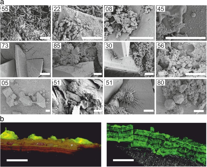
Fig. 1.
a Representative scanning electron micrographs of ureteral stent encrustations and biofilms. Numbers refer to sample IDs. Based on NGS and cultivation, bacteria visible likely include L. jensenii (ST55), G. vaginalis (ST22), S. anginosus or A. tetradius (ST08), Staphylococcus (ST45), S. epidermidis (ST85) and Corynebacterium (ST30). Further samples included structures with size and morphology of fungal cells or blood cells (ST05, ST51 and ST80). Scale bars = 5 μm. b Confocal laser scanning micrographs of SYBR Green-stained ureteral stent encrustations, shown as maximum intensity projection. Fluorescence was associated with a layered structure. Colour allocation—grey, reflection; green, SYBR Green. Scale bars = 50 μm

