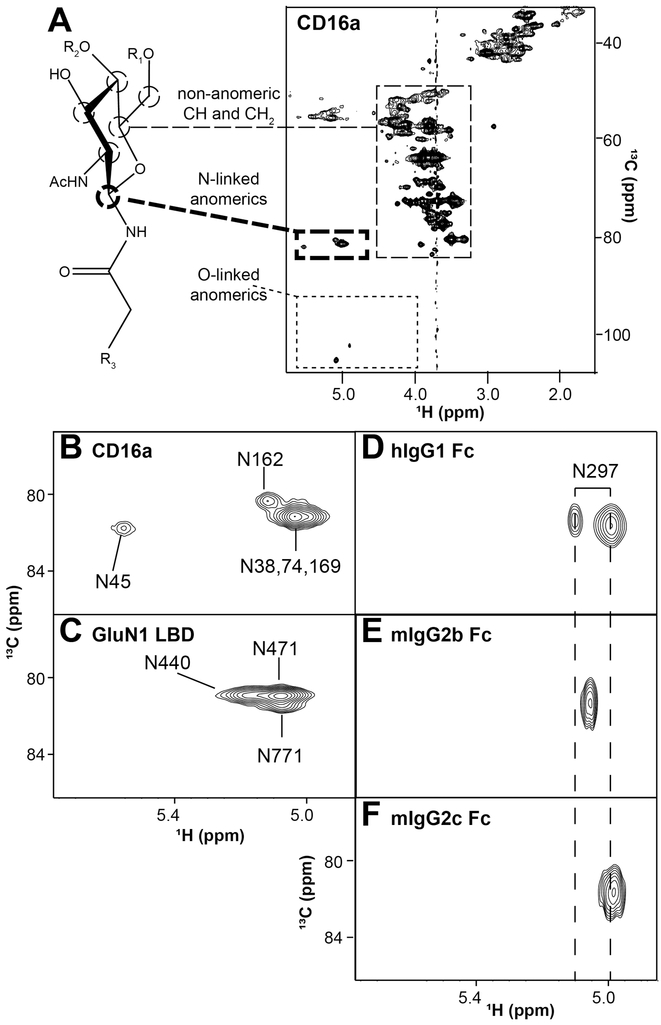
Figure 5. Proteins with 13C-labeled N-glycans provide a single, unique signal for each N-glycan.
A. A 13C-HSQC of [13C-glycan] CD16a with 5 N-glycans shows predominantly signal corresponding to carbohydrate and potentially limited scrambling to amino acids. Of the three distinct regions noted in the spectrum, crosspeaks from the anomeric asparagine-linked H1-C1 atoms are separated from the other signals, with one coherence for each N-glycan residue (expanded in B). C-F Similar analyses of four glycoproteins yields comparable results. These spectra were collected 16.4T.

