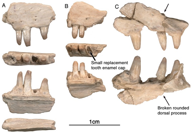
Figure 4. Arisierpeton simplex maxillae.
(A) Images of left maxillary fragment GAA 00207 in lateral, ventral, lingual, and dorsal views; (B) images of small right maxillary fragment GAA 00240 in lateral, ventral and ligual views; the ventral or occlusal view shows the presence of an unerupted tooth at the base of the brocken tooth crown, as indicated by an arrow; (C) GAA 00225-2, anterior fragment of right maxilla in labial and lingual views. Arrow points to base of rounded anterior dorsal process of maxilla.

