Figure 5.
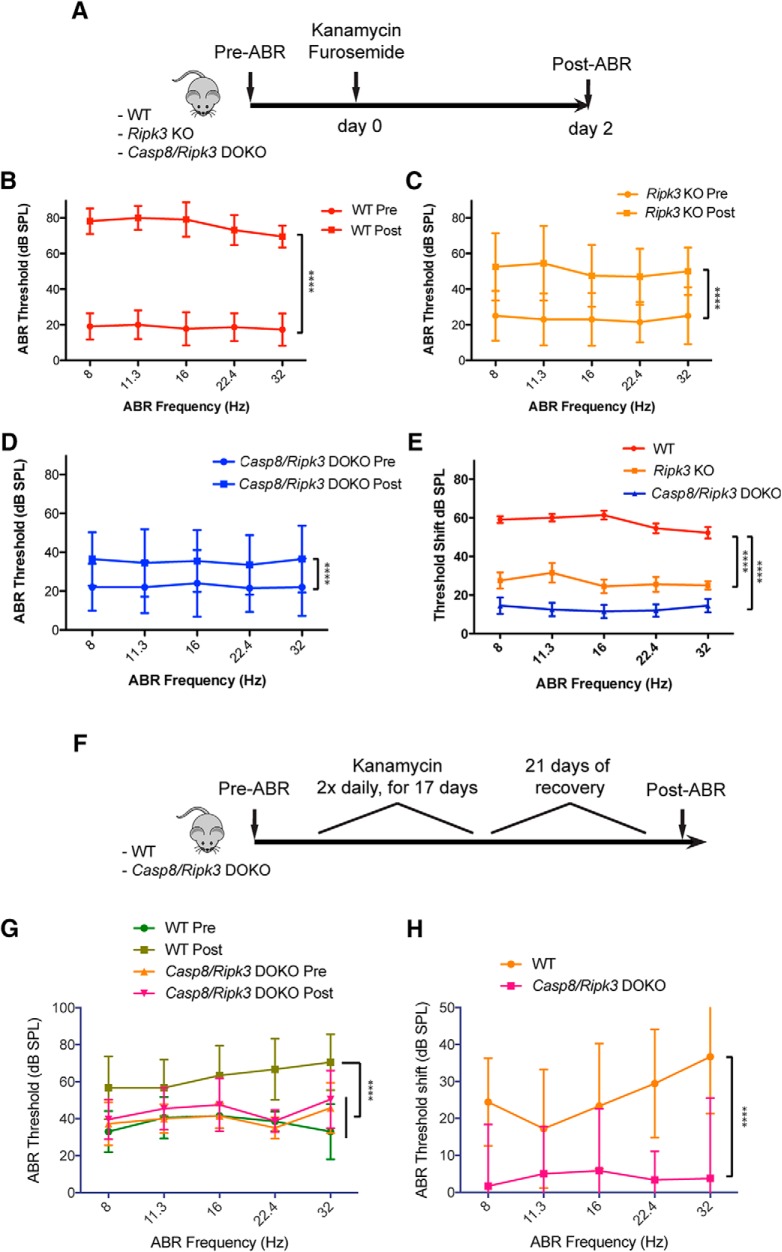
Genetic inhibition of RIPK3-mediated necroptosis and Caspase-8-mediated apoptosis alleviates kanamycin ototoxicity in vivo. A, Schematic illustration of the kanamycin/furosemide combination ototoxicity paradigm in mutant mice. B, Auditory thresholds increased ∼50–60 dB compared with pre-exposure in WT mice. C, Auditory thresholds increased ∼30 dB compared with pre-exposure in Ripk3 KO mice. D, Auditory thresholds increased ∼15–20 dB compared with pre-exposure in Casp8/Ripk3 DOKO mice. E, Degree of threshold shift at each frequency for each genotype. F, Schematic illustration of the loop diuretic-free kanamycin ototoxicity paradigm. Auditory thresholds increased significantly after kanamycin exposure in WT mice, but not in Casp8/Ripk3 DOKO mice. G, H, Plots of absolute ABR thresholds (G); and plots of the ABR threshold difference (shift) between pre-ABR and post-ABRs (H). Error bars indicate the SD. Statistical significance: ****p < 0.00001. Number of animals (n) per group: WT (n = 11, 7 females, 4 males); Ripk3 KO (n = 10, 5 females, 5 males); and Casp8/Ripk3 DOKO (n = 10, 7 females, 3 males). Number of animals (n) in the furosemide-free kanamycin protocol: WT (n = 10, 5 females, 5 males); and Casp8/Ripk3 DOKO (n = 13, 8 females, 5 males). See Table 5-1.

