Figure 1.
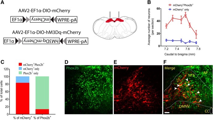
Validation of viral transfection in a Phox2b-Cre mouse line. A, Stereotaxic delivery of AAVs encoding Cre-dependent hM3Dq-mCherry or mCherry into the NTS in Phox2b-Cre mice. B, Rostrocaudal distribution of mCherry+Phox2b+ and mCherry+ NTS neurons. Cell counts were obtained in 6 coronary sections (25 μm) from each mouse (n = 6). C, Quantification of the percentage of mCherry+Phox2b+ neurons based on cell counts shown in B. The mCherry+Phox2b+ neurons accounted for ∼83% of total number of mCherry+-expressing neurons and for ∼13% of Phox2b+-expressing neurons. D–F, Most mCherry-transduced cells are Phox2b-positive. The merged photomicrograph was derived from Phox2b immunoreactivity (green, D) and mCherry (red, E). White arrows indicate the overlap of Phox2b and mCherry. Scale bar, 25 μm.

