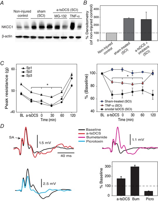
Figure 10. Mechanism of action of a‐tsDCS.

A, intrathecal injection of MG‐132 and TNF‐α blocked a‐tsDCS induced‐reduction of NKCC1 expression. Control, non‐injured and non‐stimulated mice; sham, injured but sham‐stimulated; a‐tsDCS, injured and stimulated. SCI indicates animals group with spinal cord injury. B, summary graph showing averages from different groups. C, blocking degradation of NKCC1 prevents a‐tsDCS induced reduction of spasticity. Animals with spasticity received intrathecal injection of TNF‐α or MG‐132 (n = 5) 30 min before applying a‐tsDCS and were then tested for spasticity. Left: anodal caused reduction of peak resistance that was significantly lower than baseline; however, peak resistance in response to three speeds of stretch returned exceeding baseline 2 h following the end of 20 min stimulation. Right: comparison between three groups with SCI and spasticity (speed 3 only): sham‐treated was not injected or stimulated; TNF‐α was injected and stimulated; and stimulated but not injected with TNF‐α or MG‐132. Note that these experiments were performed using the stretch device in Fig. 9 A. D, modulation of DRP by a‐tsDCS and bumetanide. DRP traces are shown on the left and top right. Bottom right: DRP was increased by a‐tsDCS and by bumetanide but reduced by picrotoxin. Bum, bumetanide; Picro, picrotoxin. [Color figure can be viewed at wileyonlinelibrary.com]
