. 2019 Apr 8;33(2):173–180. doi: 10.3341/kjo.2018.0084

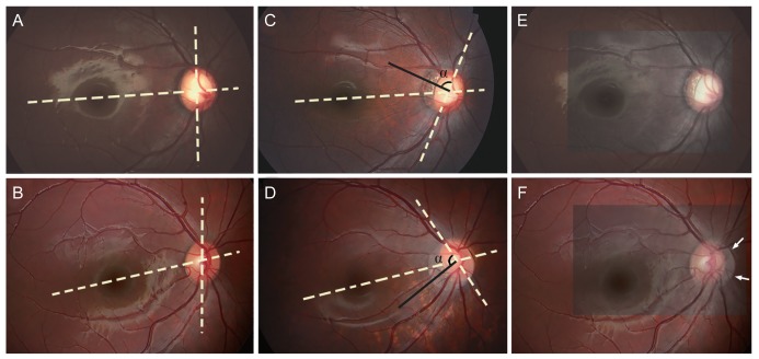
Fig. 2. Representative cases showing the development of optic-disc torsion. (A) Baseline fundus photograph of a 4-year-old girl. (B) Baseline fundus photograph of a 3-year-old girl. (C) Superior optic-disc torsion was observed 6 years later. (D) Inferior optic-disc torsion was observed 3 years later. (E) When the baseline and follow-up photographs were superimposed using the blood vessels as references, the nasal disc margin of the two photographs overlapped, indicating that the apparent torsion resulted from disc tilt on an oblique axis. (F) The disc margin of the two photographs did not overlap at any region when the two photographs were superimposed. The current disc margin is vaguely seen (arrows). (C,D) Note that the vertical disc axis (dashed line) and direction of the longest parapapillary atrophy width (black line) are approximately perpendicular.

Fig. 2