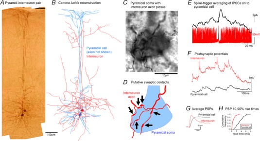
Figure 2. Rapid feedforward inhibition onto a pyramidal cell, mediated by a fast‐spiking interneuron.

A and B, photomicrograph (A) and camera lucida drawing (B) of a connected pyramidal cell–basket cell connected pair. The axonal morphology of the interneuron is consistent with descriptions of the type1 class of basket cells found in rodent visual cortex, as described by Sjöström and colleagues (Buchanan et al., 2012). C, high magnification view of the same section shown in A, showing the pyramidal soma enveloped by the interneuronal axonal plexus. D, schematic representation of A to show the locations of several putative interneuronal synaptic contacts onto the pyramidal soma. E, spike triggered averaging of the pyramidal voltage clamp recording, based on the timing of action potentials recorded in the cell‐attached interneuron, illustrating a short latency, inhibitory postsynaptic current. The example shows an almost instantaneous initial rise, that is too fast for the synaptic event, but is instead most probably caused by noise riding on top of the rapidly rising event. F, subsequent whole cell current clamp trace from the basket cell, and for comparison a similar trace from a layer 5 pyramidal cell (not synchronously recorded). Current clamp recordings were taken at resting membrane potential (E m), and since the GABAergic reversal potential (−59 mV) was close to E m (mean interneuronal E m = −60.8 ± 4.8 mV (±SEM; n = 4); mean pyramidal E m = −70.9 ± 3.6 mV), we assume that the majority of events are excitatory, although we have no direct means of separating inhibitory and excitatory PSPs in these traces. Note the large amplitude events with extremely fast kinetics in the interneuronal recording. G, average PSPs (n = 200) for a stuttering cell and the pyramidal cell. H, cumulative frequency plot of the 10–90% rise times for the two cells.
