
Figure 4.
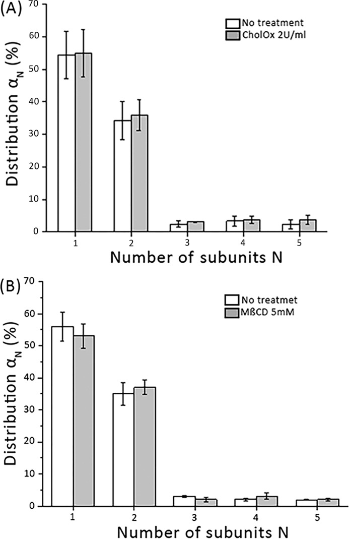
mGFP–hDAT dimeric distribution is not affected by perturbation of cholesterol. A, comparison of the dimeric mGFP–hDAT distributions in the unaltered lipid environment of the plasma membrane (white bars) and after cholesterol oxidation with cholesterol oxidase (2 units/ml for 30 min). No apparent influence of the cholesterol content is observable (n = 61 cells). B, cholesterol depletion by MβCD (5 mm for 1 h) does not affect the dimer distribution (gray bars) when compared with the unaltered lipid environment of the plasma membrane (white bars) (n = 43 cells). Error bars show the mean S.E.