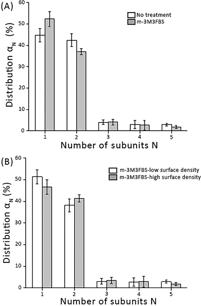
Figure 5.

mGFP–hDAT dimerization at the plasma membrane is independent of PIP2 levels. A, we enzymatically depleted PIP2 at the plasma membrane via activation of PLCγ by incubating cells for 20 min with the direct PLCγ activator m-3M3FBS (25 μm). m-3M3FBS did not yield any effect on the oligomeric distribution (gray bars) compared with the untreated cells (white bars) (n > 80 cells per experimental condition). mGFP–hDAT surface densities were similar: 35 ± 4 μm2 (white bars) and 33 ± 5 μm2 (gray bars). B, the oligomeric distribution is plotted for various mGFP–hDAT surface density (n > 40 cells) following PIP2 depletion via m-3M3FBS; mGFP–hDAT densities are ∼7 mGFP–hDAT/μm2 for low surface density and ∼40 mGFP–hDAT/μm2 for high surface density. Error bars show the mean S.E.
