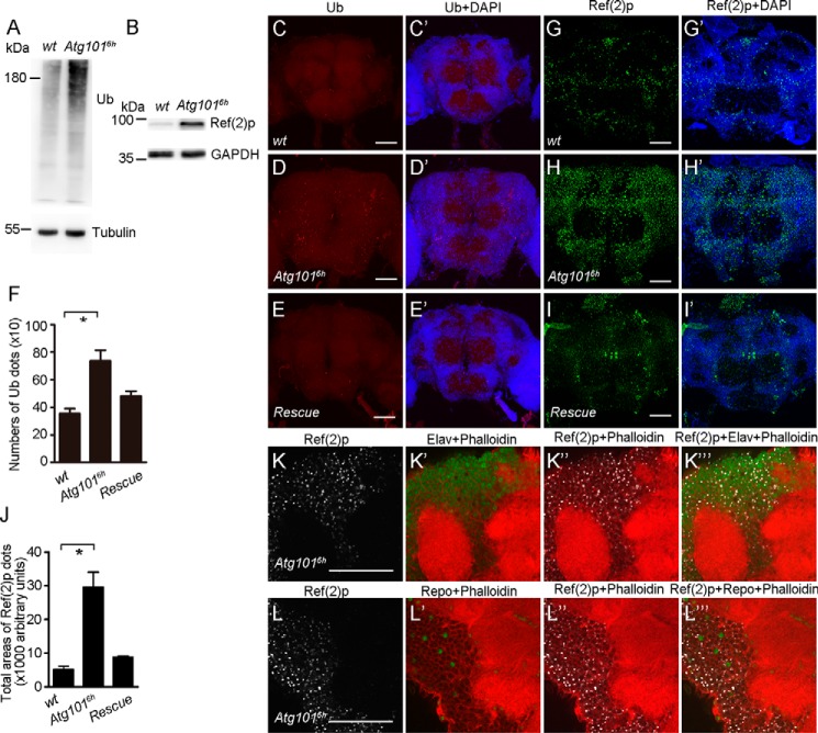
Figure 3.
Accumulation of ubiquitinated proteins and Ref(2) aggregates in Atg101 mutant brains. A, Western blotting reveals that the level of ubiquitinated proteins is increased in Atg1016h mutant fly heads. 7-day-old WT and Atg1016h mutant flies were used. Tubulin was used as a loading control. B, increased Ref(2) protein levels in Atg1016h mutant fly heads. 7-day-old WT and Atg1016h mutant flies were used. GAPDH was used as a loading control. C–E′, aggregates of ubiquitinated proteins accumulate in Atg1016h mutant brains. Shown are confocal images of Drosophila brains of 7-day-old WT and Atg1016h mutant and rescue flies stained with anti-ubiquitin antibody. Scale bars: 50 μm. F, quantification of the number of ubiquitin-positive spots in WT, Atg1016h mutant, and rescue fly brains. n = 12, 11, and 15, respectively. *, p < 0.05. G–I′, aggregates of Ref(2)p proteins accumulated in Atg1016h mutant brains. Shown are confocal images of Drosophila brains of 7-day-old WT, Atg1016h mutant, and rescue flies stained with anti-Ref(2)p antibody. Scale bars: 50 μm. J, quantification of the total areas of Ref(2)p-positive spots in WT, Atg1016h mutant, and rescue fly brains. n = 6. *, p < 0.05. K–K‴, colocalization of Ref(2)p-positive cells with Elav, a marker of neuronal nuclei. Phalloidin was used to label F-actin. Scale bars: 50 μm. L–L‴, colocalization of Ref(2)p-positive cells with Repo, a marker of glial nuclei. Phalloidin was used to label F-actin. Scale bars: 50 μm.

