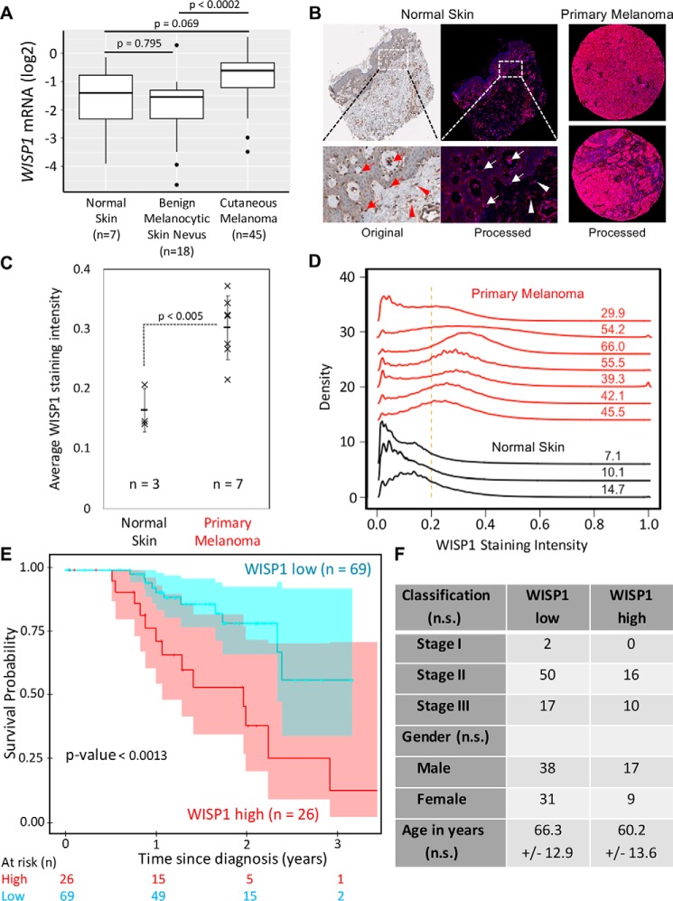
Figure 1.
WISP1 expression is increased in melanoma and is associated with reduced overall survival of patients diagnosed with primary melanoma. A, WISP1 mRNA expression in benign skin conditions (normal skin and benign melanocytic skin nevus) compared with primary melanoma. The original expression data set (GSE3189) was deposited by Talantov et al. (55). p values were calculated using analysis of variance with the post hoc Tukey honest significant difference test. B, representative original and deconvoluted color images derived from human normal skin and melanoma tissue microarray probed using a WISP1 antibody (HPA007121) and imaged using 3,3′-diaminobenzidine and stained using hematoxylin for normal skin (left) and two melanoma (right) tissue samples. Original tissue microarray images were obtained from www.proteinatlas.org3 (77). Deconvoluted intensity of WISP1 staining is shown in red, whereas the cellular structures stained using hematoxylin are shown in blue. Arrows, melanocytes in epidermis; arrowheads, fibroblasts in dermis (stroma). C, average WISP1 staining within normal skin and primary melanoma tissue samples. D, distributions in nonzero pixel intensity values of WISP1 staining for normal skin (black curves) and primary melanoma (red curves) tissue samples. Numbers, percentage of the distribution that has pixel intensity values greater than a normalized pixel intensity of 0.2. E, Kaplan–Meier estimate of overall survival of melanoma patients stratified by WISP1 transcript abundance. The original data set was from the Cancer Genome Atlas. Sample numbers and p values calculated using the Peto and Peto modification of the Gehan–Wilcoxon test are indicated. F, patient population characteristics of WISP1 high and WISP1 low groups. Statistical differences among categorical data and age were assessed using Fisher's exact test and Student's t test, respectively (n.s., p > 0.05).

