Figure 3.
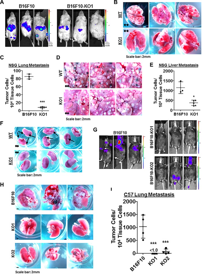
Wisp1 knockout repressed the experimental metastasis of melanoma cell line B16F10 in immunodeficient NSG mice and immunocompetent C57BL/6Ncrl mice. Experimental metastasis assays were performed in NSG mice (A–F) and C57BL/6Ncrl mice (G–I) using B16F10 and the indicated knockout cells with injection through mouse tail veins. Each group contained five duplicates (n = 5), and only mice surviving the whole experiments were analyzed at the same time for imaging, photography, and qPCR (final n ≥ 3). These experiments were repeated, and similar results were achieved. A, bioluminescence imaging performed 1 day before NSG mice were euthanized. All animals were compared with the same bioluminescence scale. B and C, tumor lung metastases (black colonies) of NSG mice as captured by photography (B) and real-time genomic qPCR (C). Quantitative tumor lung metastatic burden was assayed and presented as tumor cell number within 10,000 mouse tissue cells. D–E, tumor liver metastases (black and white nodules) of NSG mice as captured by photography (D) and real-time genomic qPCR (E). Quantitative tumor liver metastatic burden was assayed and presented as tumor cell number within 10,000 mouse tissue cells. F, tumor kidney metastases (black colonies) of NSG mice as captured by photography. G, bioluminescence imaging performed 1 day before C57BL/6Ncrl mice were euthanized. All animals were compared with the same bioluminescence scale. H–I, tumor lung metastases of C57BL/6Ncrl mice as captured by photography (H) and real-time genomic qPCR (I). Four high-resolution images for B, D, F, and H are provided as Figs. S1–S4. *, p < 0.05; **, p < 0.01; ***, p < 0.001. Error bars, S.D.

