
Figure 1.
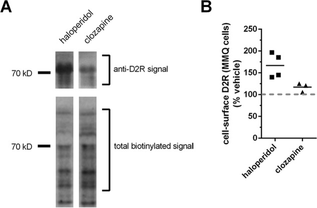
Effect of APD treatments on cell-surface levels of the D2R endogenously expressed in the pituitary-derived MMQ cell line. A, effect of APD treatment on cell-surface D2R endogenously expressed in pituitary-derived MMQ cell line. Representative lanes from Western blotting depict the effect of treatment with the indicated APDs (10 μm, 48 h) on the levels of cell-surface expression of D2R endogenously expressed in MMQ cells. Western blots of cell-surface proteins isolated by surface biotinylation of intact cells with the membrane-impermeable biotinylation reagent, EZ-LinkTM Sulfo-NHS-LC-Biotin, were probed with anti-DR antibody (top) and subsequently reprobed with HRP-conjugated streptavidin to visualize total cell-surface protein (bottom). B, quantification of cell-surface D2R in MMQ cells after treatment with the indicated APDs. Levels of endogenous cell surface D2R expression in MMQ cells visualized in A were normalized against the total cell-surface protein signal and reported as a percentage of the normalized cell-surface D2R signal in vehicle-treated cells. The signal for cell-surface D2R after treatment with haloperidol was significantly different, from both clozapine-treated (p < 0.05) and vehicle-treated (p < 0.01) cells (bar representing the mean; n = 4 for vehicle and haloperidol and 3 for clozapine; Tukey).