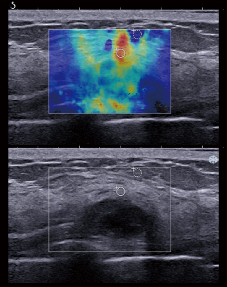
Figure 5.

A 48-year-old postmenopausal woman with a pathologically proven grade 2 invasive ductal carcinoma and luminal B subtype. The gray scale ultrasound feature showed a 1.6 cm, partially well-defined, oval hypoechoic mass, which was slightly increased in size compared with a previous US study and was assessed as BI-RADS category 4A. After applying a color overlay, SWE illustrated heterogeneous color pattern. The quantitative SWE values were 109.60 kPa for the mean elasticity, 122.95 kPa for the maximum elasticity, and 14.82 for the elasticity ratio. SWE, shear wave elastography.
