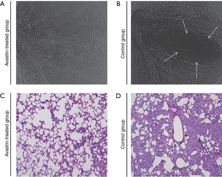
Figure 5.
Pulmonary metastasis analysis. SR phase-contrast imaging (A,B) was obtained at 20 keV with the object-to-detector distance of 1 m. The pixel size was 9 µm × 9 µm; the exposure time was 2.5 ms. (C,D), H&E-stained examination was carried in vitro. Original magnification ×200. The arrows in (B) and (D) indicated lung metastasis. Avastin-treated group (n=12), control group (n=12).

