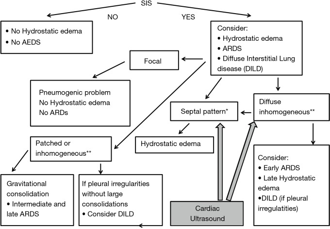
Figure 2.
Algorithmic approach for the ultrasound diagnosis of ARDS. The presence of SIS is mandatory for the identification of a parenchymal subpleural involvement. Focal SIS is indicative of a pneumogenic pathology. Septal pattern is indicative of increased hydrostatic extravascular lung water. Inhomogeneous SIS and gravitational consolidations are suggestive of established ARDS. A chronic evolution of the disease, SIS and the presence of coarse pleural irregularities indicates pulmonary fibrosis. Cardiac ultrasound can be resolutive when the differential diagnosis between late CPE and early ARDS is necessary, and mixed lung involvement (hydrostatic an fibrogenic) is possible. *, presence of separated B-lines that appear as discrete laser-like vertical hyperechoic artifacts, arising with a narrow base from the pleural line, extending to the bottom of the screen without fading, and moving synchronously with lung sliding. Typical septal B-lines show a sequence of alternating white and black (or gray) horizontal bands. **, this term refers to the inhomogeneity of the individual B-lines and to the spatial variability of the B-lines arrangement. ARDS, acute respiratory distress syndrome; SIS, sonographic interstitial syndrome; CPE, cardiogenic pulmonary edema.

