Figure 1.
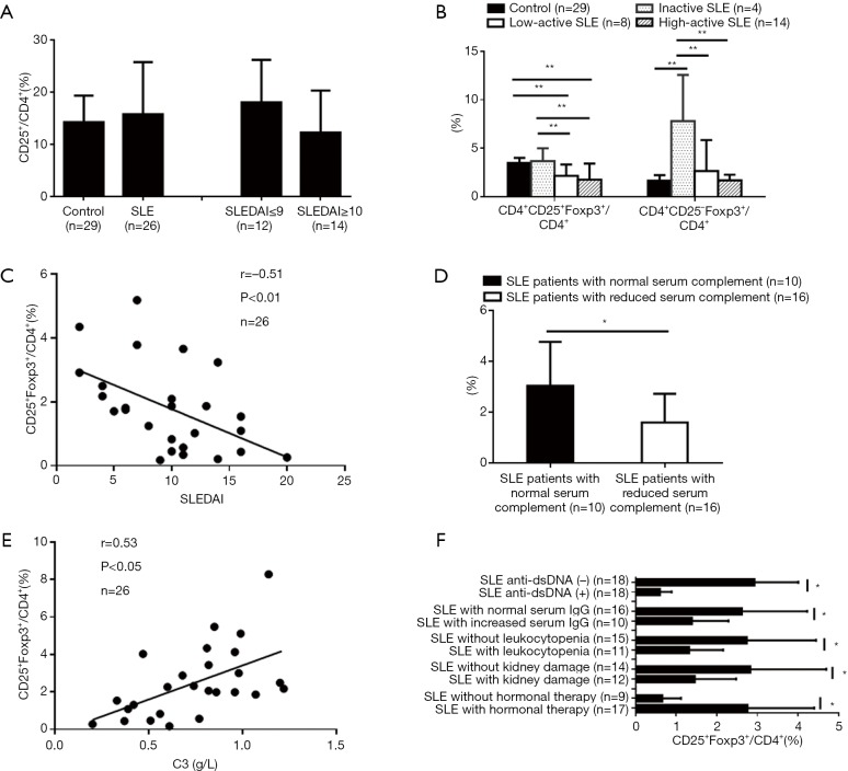
Reduction of CD4+CD25+Foxp3+Treg in SLE patients and its correlation with SLEDAI and clinical features. Flow cytometry was carried out to determine the percentages of different T cell subsets in SLE patients and controls (*, P<0.05; **, P<0.01). No changes in the percentages of the CD4+CD25+subset in CD4+T cells of SLE patients were observed compared with healthy controls (A). The percentages of the CD25+Foxp3+subset in CD4+T cells of active SLE patients decreased, and the percentages of CD25–Foxp3+ subset in CD4+T cells of inactive SLE patients increased (B). The percentage of CD25+Foxp3+subset in CD4+T cells of the patients was inversely correlated with SLEDAI (C) and also correlated with reduced serum complement (D and E), anti-dsDNA antibody positive, increased serum IgG, leukocytopenia, kidney damage, and hormonal therapy (F).

