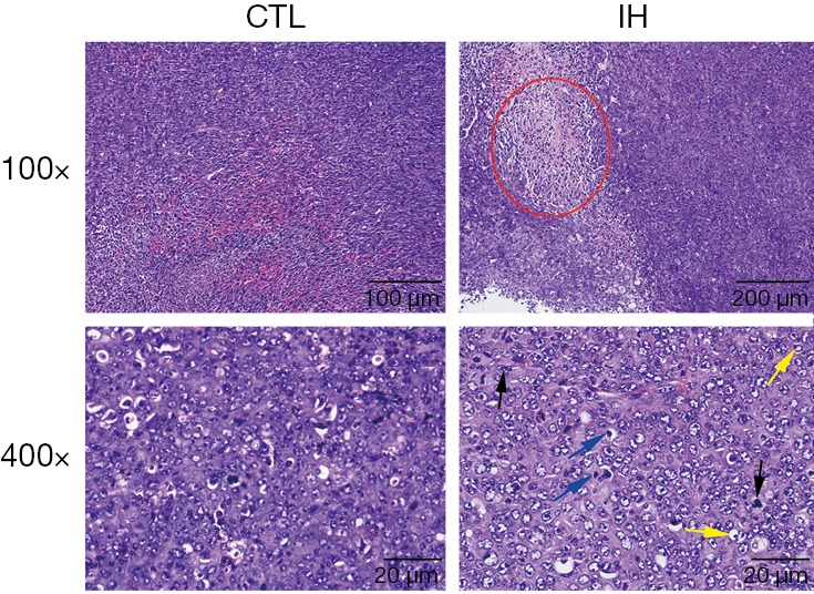
Figure 3.

Hematoxylin-eosin staining. When compared with those in the CTL group, mice in the IH group had more tumor necrosis areas (the area of the red circle). The phenomena of nuclear division (black arrows), “signet-ring cells” (yellow arrows), and deep dyeing (blue arrows) were easier to be detected in the IH group. CTL, control; IH, intermittent hypoxia.
