
Figure 2.
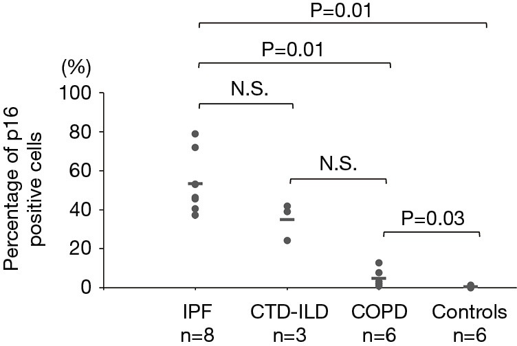
Percentage of p16-positive cells in the IPF, CTD-ILD, COPD and control groups. The horizontal bars indicate average values. The Kruskal-Wallis test was used. IPF, idiopathic pulmonary fibrosis; CTD-ILD, connective tissue disease-associated interstitial lung disease; COPD, chronic obstructive pulmonary disease; N.S., not significant.