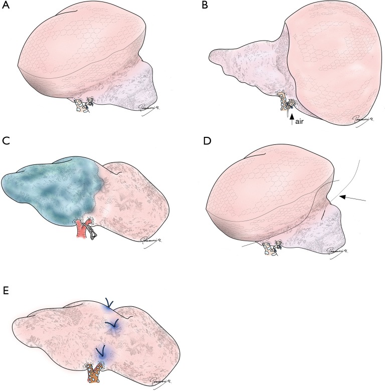
Figure 6.
Different techniques to identify intersegmental planes and/or resection lines. (A) Conventional inflation/deflation with the target segment deflated; (B) modified inflation/deflation with the target segment inflated; (C) intravenous indocyanine green injection with the pulmonary artery supplying the target segment temporarily clumped; (D) combination of a localization technique (e.g., hookwire) with a technique to identify intersegmental planes (inflation/deflation technique, in this figure). The interrupted line indicates the resection line extended into an adjacent segment (i.e., extended segmentectomy). The arrow indicates a hookwire placed under the guidance of computed tomography; (E) virtual-assisted lung mapping or bronchoscopic multi-spot dye marking. “Standing stitches” are placed, in this figure, for easier identification during stapling. See reference (23) for details.

