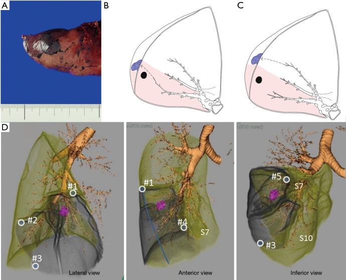
Figure 7.
The principle of VAL-MAP-assisted segmentectomy. (A) A good dye mark in VAL-MAP remains in a single secondary lobule without disseminating across the intersegmental plane. (B,C) Marks placed close to the intersegmental line bordered by the intersegmental veins. The target segment is shown as a shadowed area. A mark is placed from a bronchus located in the target segment (B) or from an adjacent segment (C). (D) A case of S8+9 anatomical segmentectomy of the right lower lobe. Corresponding three-dimensional images showing five marks (#1–#5) placed by VAL-MAP along the resection lines, particularly at the corner. The target segments are shown in gray. The figure is cited from reference (8) with permission. VAL-MAP, virtual-assisted lung mapping.

