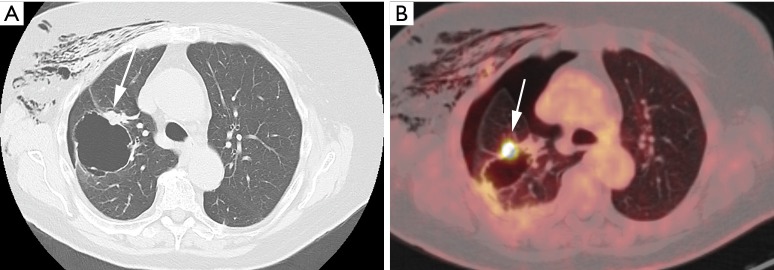
Figure 1.
An 87-year-old woman presented at the emergency department with acute chest pain and respiratory distress. Plain radiograph (not shown) depicted a large right-sided pneumothorax. Axial CT in lung window setting (A) after chest tube drainage shows a large thin-walled cystic airspace in the right upper lobe with exophytic solid nodule (white arrow). Also note the residual subcutaneous emphysema in the right breast and chest wall. 18F-FDG-PET examination showed (B) a high uptake in the exophytic solid nodule (white arrow). Histopathologic examination after right upper lobe lobectomy revealed a 1.5 cm poorly differentiated adenocarcinoma with high-grade dysplasia and focal areas of adenocarcinoma in situ in the cyst wall. CT, computed tomography; 18F-FDG-PET, 18F-fluorodeoxyglucose positron emission tomography.

