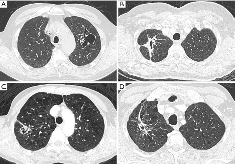
Figure 10.
Mimickers of lung cancer associated with cystic airspaces. (A) A 73-year-old man with a previous history of laryngeal squamous cell carcinoma presented on CT with a persistent large cystic lesion in the left upper lobe. The lesion showed an overall thin wall with some focal asymmetries and nodular components. Lobectomy was performed in acute setting of massive hemoptysis. Histopathologic examination confirmed diagnosis of a bronchogenic cyst with no signs of malignancy; (B) axial CT-image in lung window setting in a 52-year-old woman with previous history of massive pulmonary embolism showing an ill-defined area of consolidation with central cystic airspace. Comparison with older CT-imaging studies confirmed that this was a large involuting pulmonary infarction; (C) a 69-year-old woman presented to the thoracic surgeon with a malignant nodule (not shown) in the right upper lobe. Axial CT-image showed in the same lobe a more nodular, relatively well-defined area of consolidation with confluent cystic airspaces. Histopathologic examination after right upper lobe lobectomy showed no signs of malignancy but diagnosis of fungal infection, aspergilloma; (D) a 62-year-old man with no previous medical history presented with hemoptysis. Axial CT showed an ill-defined spiculated area of consolidation in the right upper lobe with central cystic airspace and small endophytic mural nodule. Histopathologic examination after lobectomy showed no signs of malignancy, but a cavitated lesion with inflammatory granulation tissue and a central small lung ball resulting from infection by Candida. CT, computed tomography.

