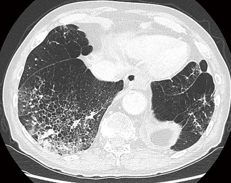
Figure 11.

A 79-year-old man with persistent cough was referred to the radiology department for chest CT-imaging. Axial image in lung window setting at the lung base shows extensive emphysema with confluent areas of consolidation in the right lower lobe. Due to the underlying emphysematous changes, the superimposed consolidation has a honeycombing-like appearance and some areas have a more nodular aspect. Since the patient also had fever, findings were considered to be infectious. Follow-up was advised and showed involution of the consolidation. Correlation with clinical findings and close monitoring of evolution is mandatory in these cases. CT, computed tomography.
