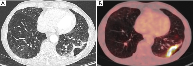
Figure 12.
A 74-year-old man with history of COPD presented to the pulmonologist with recurrent episodes of infection. Chest CT (A) showed extensive emphysematous changes in both lungs, with more prominent bullous changes or cystic airspace in the subpleural region of the left lower lobe with associated bandlike consolidation. Since this finding did not significantly changed during follow-up (no increase, but also no decrease) and bronchoscopy and BAL showed no abnormalities, further work-up was done. 18F-FDG-PET (B) showed an intense uptake in the consolidation. In combination with the evolution, a malignant cause was suggested. Histopathologic examination after surgery, revealed an invasive adenocarcinoma with areas of lepidic growth. COPD, chronic obstructive pulmonary disease; CT, computed tomography; BAL, bronchoalveolar lavage; 18F-FDG-PET, 18F-fluorodeoxyglucose positron emission tomography.

